Camera Pill Endoscopy: Swallowing a Tiny Camera Instead of a Colonoscopy
Last reviewed by staff on May 23rd, 2025.
Introduction
Camera pill endoscopy, also called capsule endoscopy, is a method for viewing the inside of the gastrointestinal (GI) tract using a small camera that is swallowed. This camera is contained within a pill-shaped capsule, which transmits real-time images of the digestive tract to a recording device. Clinicians can review these images to detect conditions such as bleeding, inflammation, ulcers, and tumors.
This procedure is considered minimally invasive. It often does not require sedation, and it typically results in fewer complications than a standard colonoscopy or endoscopy. The camera pill is usually disposable, meaning it passes naturally through the body without needing extraction. Patients generally report less discomfort with camera pill endoscopy. Because of these advantages, many clinicians view it as a significant development in gastrointestinal diagnostics. This article describes how camera pill endoscopy works, the preparation involved, its potential benefits and limitations, and how it compares with a traditional colonoscopy.
What Is Camera Pill Endoscopy?
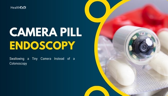
Camera pill endoscopy is a diagnostic procedure that uses a capsule roughly the size of a large multivitamin. Inside the capsule, there is:
- A miniature camera
- A light source (often LEDs)
- A transmitter and battery
When a patient swallows this pill, the device travels through the digestive tract, capturing and sending images to a receiver worn by the patient—often on a belt or harness. After a typical transit time that may last eight to 12 hours, the battery expires, and the capsule is eventually passed in a bowel movement. During those hours, thousands of images are captured. Clinicians then examine the image data for any signs of abnormality.
Key Innovations
- Wireless Technology: The transmitter within the capsule sends data to an external receiver without wires. This allows patients freedom to move around during the procedure.
- Disposable Nature: Most camera capsules are designed for one-time use. They pass through the digestive system without the need for retrieval.
- Multi-Section Coverage: Traditional procedures like standard endoscopies or colonoscopies typically focus on the upper GI tract or the colon. Camera pill endoscopy can visualize the small intestine, an area that is more difficult to access with traditional equipment.
How Does Camera Pill Endoscopy Work?
Swallowing the Capsule
The procedure generally begins in the morning. Patients swallow the capsule with water, much like taking a regular pill. After swallowing, the external receiver starts capturing signals transmitted by the camera.
Image Transmission and Recording
As the capsule moves along, the camera snaps pictures—often several frames per second. These images are transmitted wirelessly to the receiver. The receiver often shows indicator lights to confirm image recording status.
Transit Through the GI Tract
Within the small intestine, the pill is carried by natural digestive motions. It captures images in multiple sections:
- Esophagus: The capsule passes quickly, so fewer images of this segment are gathered.
- Stomach: The capsule floats and churns around in gastric fluids, potentially capturing additional details of the stomach lining.
- Small Intestine: This region is notoriously challenging to assess with traditional scopes, but the camera pill usually captures thousands of images in this lengthy section.
- Colon: After passing the small intestine, the capsule enters the large intestine. By this time, the battery life may vary, so some images may or may not be collected in the colon, depending on how quickly the capsule travels.
Data Extraction
After the recommended recording time—about eight to 12 hours—the patient returns the receiver to the clinic. The recorded images are downloaded to a computer system. A specialist then reviews the images using specialized software.
Capsule Exit
Once the capsule finishes its journey, it exits the body during normal bowel movements. Patients typically do not feel the capsule passing.
Uses of Camera Pill Endoscopy in GI Diagnostics
Camera pill endoscopy helps clinicians identify a range of gastrointestinal conditions:
- Small Bowel Bleeding
- Often, unexplained GI bleeding originates in the small intestine. Capsule endoscopy can find sources of bleeding such as ulcers or arteriovenous malformations.
- Often, unexplained GI bleeding originates in the small intestine. Capsule endoscopy can find sources of bleeding such as ulcers or arteriovenous malformations.
- Inflammatory Bowel Disease (IBD)
- Crohn’s disease affects the small intestine in many cases. The capsule can detect inflamed areas, ulcers, or narrowing (strictures) that might go unnoticed with other methods.
- Crohn’s disease affects the small intestine in many cases. The capsule can detect inflamed areas, ulcers, or narrowing (strictures) that might go unnoticed with other methods.
- Polyps and Tumors
- Polyps or tumors in the small intestine can be difficult to visualize with conventional scopes. The capsule may reveal these growths and help clinicians decide on further investigation or therapy.
- Polyps or tumors in the small intestine can be difficult to visualize with conventional scopes. The capsule may reveal these growths and help clinicians decide on further investigation or therapy.
- Celiac Disease
- Celiac disease can cause changes in the lining of the small intestine. Capsule images can show villous atrophy or related findings.
- Celiac disease can cause changes in the lining of the small intestine. Capsule images can show villous atrophy or related findings.
- Esophageal Disorders (limited)
- Certain capsules are designed specifically for the esophagus. They may help detect Barrett’s esophagus or esophageal varices.
- Certain capsules are designed specifically for the esophagus. They may help detect Barrett’s esophagus or esophageal varices.
- Colonic Evaluation
- While the procedure is often aimed at the small intestine, some capsules are designed to capture images in the colon as well. However, for direct intervention (like a biopsy), a standard colonoscopy is still needed.
- While the procedure is often aimed at the small intestine, some capsules are designed to capture images in the colon as well. However, for direct intervention (like a biopsy), a standard colonoscopy is still needed.
Potential Benefits and Limitations
Benefits
- Minimally Invasive
- Camera pill endoscopy is usually well tolerated. It does not involve a long endoscope tube. This reduces discomfort and anxiety for many patients.
- Camera pill endoscopy is usually well tolerated. It does not involve a long endoscope tube. This reduces discomfort and anxiety for many patients.
- No Sedation Required
- Most patients remain awake and go about their day. They do not need anesthesia, which lowers risks related to sedation.
- Most patients remain awake and go about their day. They do not need anesthesia, which lowers risks related to sedation.
- Small Intestine Visualization
- Camera pills can capture images of areas that cannot be easily reached by a traditional endoscope.
- Camera pills can capture images of areas that cannot be easily reached by a traditional endoscope.
- Reduced Procedure Time in Clinic
- The actual clinic visit is usually short. A patient swallows the capsule, puts on a receiver, and can resume daily activities. The main time investment is reviewing the images afterward, but that does not involve patient presence.
- The actual clinic visit is usually short. A patient swallows the capsule, puts on a receiver, and can resume daily activities. The main time investment is reviewing the images afterward, but that does not involve patient presence.
- Potential for Early Diagnosis
- Because the capsule can detect subtle lesions or bleeding sites, it can lead to earlier diagnosis and targeted treatment.
- Because the capsule can detect subtle lesions or bleeding sites, it can lead to earlier diagnosis and targeted treatment.
Limitations
- Lack of Therapeutic Intervention
- Unlike a traditional colonoscope or endoscope, a capsule cannot deliver therapy (e.g., remove polyps, take direct biopsies, or stop bleeding). Any abnormal finding on capsule endoscopy may still require a follow-up procedure.
- Unlike a traditional colonoscope or endoscope, a capsule cannot deliver therapy (e.g., remove polyps, take direct biopsies, or stop bleeding). Any abnormal finding on capsule endoscopy may still require a follow-up procedure.
- Battery Life Constraints
- If the capsule moves slowly, the battery may run out before reaching the colon. This can lead to incomplete examinations.
- If the capsule moves slowly, the battery may run out before reaching the colon. This can lead to incomplete examinations.
- Potential for Retention
- Although rare, the capsule can get stuck, especially if strictures or other obstructions are present. If this happens, surgical or endoscopic retrieval might be necessary.
- Although rare, the capsule can get stuck, especially if strictures or other obstructions are present. If this happens, surgical or endoscopic retrieval might be necessary.
- Image Interpretation Complexity
- Reviewing thousands of images requires time and expertise. Clinicians may need specialized training. Even with advanced software, interpretation can be challenging.
- Reviewing thousands of images requires time and expertise. Clinicians may need specialized training. Even with advanced software, interpretation can be challenging.
- Incomplete Examination of the Colon
- Camera pills are valuable in viewing the small intestine. However, they might not deliver the same in-depth view of the entire colon as a traditional colonoscopy.
- Camera pills are valuable in viewing the small intestine. However, they might not deliver the same in-depth view of the entire colon as a traditional colonoscopy.
Comparison with Traditional Colonoscopy
A traditional colonoscopy uses a flexible, camera-equipped tube that is inserted through the rectum. It provides high-quality, real-time images of the colon. In addition, clinicians can perform interventions during colonoscopy, such as removing polyps or taking biopsies. However, some patients find colonoscopy to be uncomfortable. Sedation is usually given, which may introduce additional risks or limit the patient’s ability to drive on the same day.
On the other hand, camera pill endoscopy involves swallowing a small camera. It usually requires less sedation or none at all. The images captured cover a broader range of the GI tract, although the focus is often on the small intestine. Below is a simplified comparison:
| Aspect | Camera Pill Endoscopy | Traditional Colonoscopy |
| Invasiveness | Swallowed capsule, minimal insertion | Colonoscope inserted rectally |
| Sedation | Often none | Usually required |
| Coverage | Primarily small intestine, sometimes partial colon | Focuses on colon (large intestine) |
| Therapeutic Options | None (diagnostic only) | Biopsies, polyp removal, bleeding control |
| Time Commitment | Short clinic visit, but 8+ hours wearing receiver | Usually 30–60 minutes under sedation |
| Comfort | Less discomfort, can resume normal activities | May cause discomfort, sedation downtime |
| Battery/Power Limits | Around 8–12 hours of image capture | Not applicable, real-time endoscopy |
| Risk of Retention | Possible in strictures | Not applicable in the same way |
Steps and Preparation for the Procedure
Pre-Procedure Assessment
- Medical History: Clinicians check for any history of bowel obstructions, strictures, or prior surgeries that could affect capsule passage.
- Medication Review: Certain medications may need adjustments. For instance, iron supplements can affect imaging clarity.
Bowel Preparation
- Dietary Restrictions: Some clinicians recommend a clear-liquid diet or fasting for eight to 12 hours before swallowing the capsule.
- Laxatives: In certain cases, a bowel cleanse is prescribed to improve visibility in the small intestine. This is less extensive than a full colonoscopy prep but still important for image clarity.
Swallowing the Capsule
- Timing: The capsule is often swallowed in the morning. Patients are instructed to wear the external receiver immediately.
- Positioning: Most patients swallow the capsule with water while standing upright to help it pass down smoothly.
During the Procedure
- Normal Activities: Patients can usually walk, sit, and engage in light tasks. Some are advised to avoid strenuous exercise or activities that could dislodge the receiver or sensor array.
- Diet: Some protocols allow a light meal a few hours after swallowing the capsule. This can vary depending on clinical instructions.
Post-Procedure
- Receiver Return: After the designated monitoring period (eight to 12 hours), the patient returns the receiver.
- Data Download: The clinic staff transfers the recorded images onto a computer for review.
- Capsule Passage: The capsule exits naturally in a bowel movement, usually within 24 hours. The patient typically does not need to retrieve it.
Risks and Complications
Although camera pill endoscopy is considered safe, a few risks are possible:
- Capsule Retention
- This can occur if there is a narrowing, obstruction, or severe inflammation. In rare cases, removal may require endoscopic or surgical intervention.
- This can occur if there is a narrowing, obstruction, or severe inflammation. In rare cases, removal may require endoscopic or surgical intervention.
- Inaccurate or Incomplete Data
- A slow transit time can cause the battery to run out before full passage. Also, if the bowel is not sufficiently cleansed, images may be unclear.
- A slow transit time can cause the battery to run out before full passage. Also, if the bowel is not sufficiently cleansed, images may be unclear.
- Misinterpretation of Images
- Reviewing thousands of images demands skill. Small lesions might be overlooked if the dataset is large or if bowel contents obscure part of the camera’s view.
- Reviewing thousands of images demands skill. Small lesions might be overlooked if the dataset is large or if bowel contents obscure part of the camera’s view.
- Allergic Reactions (rare)
- The capsule’s materials are typically biocompatible, and true allergic reactions are uncommon.
- The capsule’s materials are typically biocompatible, and true allergic reactions are uncommon.
Who Can Benefit from Camera Pill Endoscopy?
Patients with Suspected Small Bowel Disease
Many individuals who experience unexplained GI bleeding or chronic anemia might benefit from capsule endoscopy. Traditional methods sometimes fail to locate bleeding sources in the small intestine.
Those with Crohn’s Disease
Crohn’s disease often affects the small bowel. Capsule endoscopy can visualize inflamed areas that remain unreachable by standard scopes.
Patients Who Avoid Standard Endoscopy
Some patients are at higher risk for sedation complications or are anxious about sedation. Capsule endoscopy might be an alternative for these individuals.
Post-Surgical Cases
Patients who have had certain surgeries might have anatomical changes in the GI tract. A camera pill can sometimes navigate through these altered pathways for a basic diagnostic overview.
Screening in Special Situations
Though not the first choice for standard colon screening, some high-risk patients or individuals who cannot undergo standard colonoscopy might be guided to consider a specialized colonic capsule.
Future Advances in Endoscopic Technology
Camera pill technology continues to develop. Engineers are refining battery life, image resolution, and data transfer rates. Some experimental capsules are being designed with sensors to detect pH, temperature, or chemicals that may indicate bleeding. Research also focuses on:
- Motorized Capsules
- Prototypes include tiny motors or magnets allowing clinicians to control the capsule’s position.
- Prototypes include tiny motors or magnets allowing clinicians to control the capsule’s position.
- Real-Time Viewing
- Some models are exploring live streaming of images, letting clinicians observe the capsule’s journey without delay.
- Some models are exploring live streaming of images, letting clinicians observe the capsule’s journey without delay.
- Artificial Intelligence (AI) Integration
- New software aims to highlight suspicious lesions automatically. This could reduce the time required for image review and possibly increase diagnostic accuracy.
- New software aims to highlight suspicious lesions automatically. This could reduce the time required for image review and possibly increase diagnostic accuracy.
- Biopsy Capabilities
- Though still experimental, efforts are underway to allow capsules to collect tissue samples. This would greatly expand diagnostic utility.
- Though still experimental, efforts are underway to allow capsules to collect tissue samples. This would greatly expand diagnostic utility.
These innovations could bring camera pill endoscopy closer to the diagnostic and therapeutic power of a traditional colonoscopy without causing the same level of discomfort.
Conclusion
Camera pill endoscopy represents a major advancement in visualizing the gastrointestinal tract, especially the small intestine. By swallowing a small capsule equipped with a camera, patients can enjoy a minimally invasive experience that does not usually require sedation.
This method can detect ulcers, bleeding, polyps, or inflammatory conditions that might otherwise evade diagnosis.
Although capsule endoscopy cannot replace all functions of a standard colonoscopy—particularly intervention, polyp removal, or direct biopsy—it is well-suited for identifying problems deep in the small intestine.
Its benefits include a relatively simple preparation, minimal patient discomfort, and wide coverage of the GI tract. With ongoing research and improvements in design, camera pill endoscopy may become an increasingly important tool in gastrointestinal diagnosis. It offers a new perspective on detecting conditions that were once difficult to visualize.
References
- Iddan G, Meron G, Glukhovsky A, Swain P. Wireless capsule endoscopy. Nature. 2000;405(6785):417.
- Delvaux M, Gay G. Capsule endoscopy: Technique and indications. Best Pract Res Clin Gastroenterol. 2008;22(5):813–837.
- Liao Z, Hou X, Lin-Hu EQ, Sheng JQ, Ge ZZ, Jiang B, et al. Accuracy of capsule endoscopy in the identification of small bowel diseases. Gastrointest Endosc. 2010;71(2):280–286.
- Adler DG, Chand B, Conway JD, Diehl DL, Fennerty MB, Kantsevoy SV, et al. Capsule endoscopy of the small bowel. Gastrointest Endosc. 2008;68(4):621–623.
- Spada C, Koulaouzidis A, Hassan C, Pennazio M, Adler S, Ge Z, et al. Colon capsule endoscopy: Current status and future directions. World J Gastroenterol. 2021;27(12):1151–1161.
- Food and Drug Administration (FDA). Wireless capsule endoscopy devices. FDA.gov. Published 2020.
- American College of Gastroenterology (ACG). Guidelines for small bowel endoscopy. Am J Gastroenterol. 2015;110(4):452–464.
- Barkin JS, Friedman S. Wireless capsule endoscopy requiring surgical intervention: The world’s experience. Am J Gastroenterol. 2002;97(9):2162–2165.
- Cave D, Legnani P, de Franchis R, Lewis BS. ICCE consensus for capsule retention. Endoscopy. 2005;37(10):1065–1067.
- Rondonotti E, Pennazio M, Toth E, Koulaouzidis A. Small-bowel capsule endoscopy: A ten-point contemporary review. World J Gastroenterol. 2021;27(40):6796–6814.