Pfeiffer syndrome
Last reviewed by Dr. Raj MD on January 12th, 2022.
What is Pfeiffer syndrome?
This is a rare genetic disorder that affects the shapes of the face and head of patients and is characterized by the premature fusion of certain bones of the skull. This affects the shape of their face and head and will prevent the further growth of the skull. It can also affect the bones in the feet and hands. Around the world it affects approximately one in one hundred thousand births. It is named after Rudolf Arther Pfeiffer, a German geneticist in 1964.
Types of Pfeiffer syndrome
There are three types of Pfeiffer syndrome, which are classified according to how severe their symptoms are. The three types include:
- Type 1 – this form of Pfeiffer syndrome is characterized by the premature fusion of the cranial sutures but does not affect their intelligence. With this type there is a survival rate that is significant. When a person has this type they will normally have various finger and toe abnormalities along with recessed cheekbones. This type is also known as the classic Pfeiffer syndrome.
- Type 2 – with this form of Pfeiffer syndrome the main characteristic is the abnormal shape of their skull which looks like a “cloverleaf” which happens because of the fusion of the cranial sutures which are extensive. Having a cloverleaf shaped skull normal brain growth can be hampered, which in turn can cause mental retardation and neurological problems.
- Type 3 – with this form of Pfeiffer syndrome the person will have symptoms that are similar to type 2 but the person does not have the cloverleaf shape of the skull.
Symptoms
The symptoms of Pfeiffer syndrome will vary from infant to infant but the milder symptoms are seen with Type 1. Some of the common symptoms that are seen can include:
- Abnormal skull that appears tower-shaped.
- High broad forehead
- Short wide thumbs
- Toes that are big
- Short partially developed toes and fingers.
- Hands and feet that are webbed.
- Problems with speech
- Deformities of the face because of underdevelopment.
- Wide, low set, down slanting eyes or bulging eyes.
- Upper jaw bone is small.
- Nasal bridge is low set and can have a beaked nose.
- Deformation of the larynx, the voice box, and the part that connects the larynx with the lungs, called the pharynx.
- Having an outward projecting chin.
- Visual axes that are different.
- Loss of hearing
- There is a malformation of the passage between the nose and pharynx.
- Having oral deformities like a high palate.
- Having an abnormal formation of their teeth.
- Having a fixed elbow joint which is seen occasionally.
- Having a cleft palate.
- Due to airway complications they can have respiratory distress.
- Due to tracheal deformities they may have a hoarse voice.
Pfeiffer syndrome Causes
The cause of Pfeiffer syndrome is genetic and is associated with the Fribroblast growth factor receptors one and two. These receptors are important for normal, healthy bone development. In Pfeiffer syndrome these growth factors mutate and affect the bones in the head. This mutation will normally happen before the child is born in the mother’s uterus causing the head to be unable to grow in the normal way. Pfeiffer syndrome is considered a rare autosomal dominantly inherited disorder. At this time there are no known ways to prevent this genetic disorder from developing.
Diagnosis
In order to diagnosis Pfeiffer syndrome it will usually take a team of orthopedists, ophthalmologists, and medical geneticists to make the diagnosis. A through physical examination is also necessary to help in making the diagnosis. The physicians will also do a variety of diagnostic tests such as regular x-rays, MRI (Magnetic Resonance Imaging) or a CT scan (computer tomography scan) to help detect the fusion of bone structures and coronal cranial structures that are untimely. Although they do not usually do prenatal diagnosis to detect Pfeiffer syndrome there have been some cases where they did use this diagnostic procedure to identify the cloverleaf skull deformity in an unborn child. They can also do a proper three-dimensional ultrasound to help diagnosis Pfeiffer syndrome without the cloverleaf skull being present.
Treatment
The treatment that is used depends on how severe the symptoms are and the age of the onset of Pfeiffer syndrome. The treatment that is usually used consists of a variety of surgeries and medications to help correct the deformities and manage the symptoms. There will usually be a team of medical experts such as neurosurgeons, dentists, plastic surgeons, Ear, Nose, and Throat specialist, and more involved in the treatment of the infant. The surgeries that will be involved in the treatment include removing and replacing the deformed skull bone. This will help to relieve the pressure on their brain. In order to help manage their breathing and eating difficulties they may need to have a tracheotomy and gastrostomy surgery. They may also need surgery to repair their webbed hands and feet when they are older. To correct the facial deformities they may need correction and cosmetic reconstruction surgery.
It is necessary to have a pro-active treatment plan along with a long-term surgical plan in order for them to maintain a normal life. Usually the abnormalities of their legs cannot be corrected. They can surgically partially correct or modify the abnormal elbow joints.
Pfeiffer syndrome Pictures
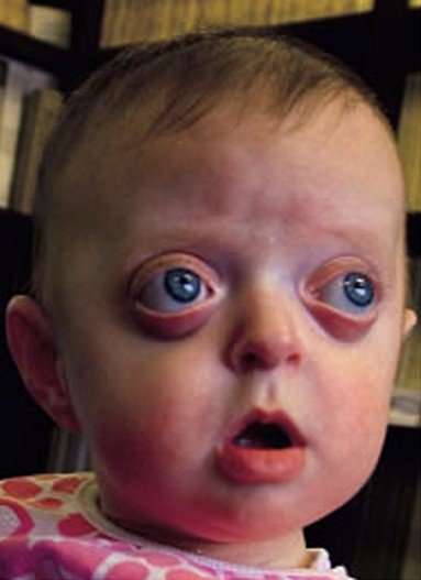 Pfeiffer syndrome (Type 1)
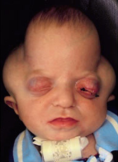 Pfeiffer syndrome (Type 2)
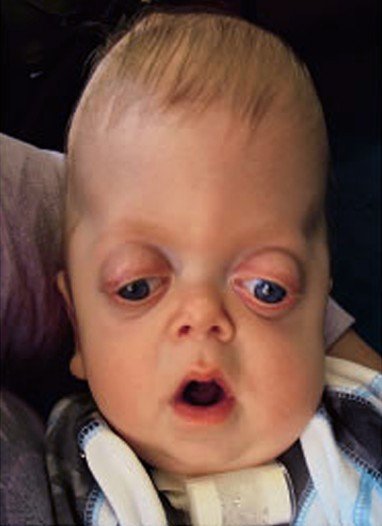 Pfeiffer syndrome (Type 3)