Osteogenesis Imperfecta Type IV
Last reviewed by Dr. Raj MD on January 12th, 2022.
Osteogenesis imperfecta is a form of genetic disease in which the bone of the patient breaks easily. For this reason, Osteogenesis imperfecta is called brittle bone disease. It is associated with a malfunctioning of one of the genes that make protein (type 1 collagen).
This protein is a primary component of the connective tissues in the bones. It also aids in the formation of ligaments, white outer tissue of the eyeballs, and teeth. Because of the defective gene, the production of type 1 collagen is impaired. This has resulted to weak bones that tend to break easily. (1, 2)
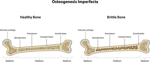
Image 1: An image comparison of a healthy bone and a bone with osteogenesis imperfecta/brittle bone disease.
Photo Source: ghr.nlm.nih.gov
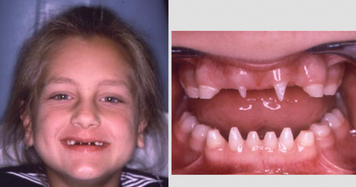
Photo 3: A clinical manifestation of a female patient with osteogenesis imperfecta.
Is it a genetic disease?
Most cases of Osteogenesis imperfecta is caused by a defective gene from any of the parents. There are instances though when none of the parents have OI. The reason for OI is spontaneous mutation of the gene. It came to a point when the gene is no longer functioning correctly. (2)
What are the clinical manifestations?
- The patient’s teeth are brittle
- Short stature
- Hearing loss
- Breathing-related problems
- Triangular shaped face
- Deformity of the bone like scoliosis or bowed legs (2, 3, 4)
Osteogenesis Imperfecta Diagnosis
The first sign of osteogenesis imperfecta is a bone that easily breaks even with no or little force. To confirm the diagnosis, clinical studies and laboratory work should be done. These include the following:
- Physical examination – The doctor will perform a thorough physical examination and paying particular attention to the eyes and teeth.
- X-ray – This procedure is done to clearly visualize the bones of the patient and see whether there are fractures or bone malformation.
- Blood/tissue sample – A sample of blood or tissue is needed for genetic testing.
- Ultrasound – This procedure helps detect severe cases of osteogenesis imperfecta, especially during pregnancy. (4, 6, 7, 8)
What causes osteogenesis imperfecta?
Osteogenesis imperfecta is a genetic disorder. It can be inherited from the parents. One of the genes is not working the way it supposed to be. The gene that causes osteogenesis imperfecta affects the formation of collagen. It is the collagen that makes the bone strong. If the genes are not performing the right way, the collagen level is altered causing brittle and weak bones.
Types of osteogenesis imperfecta
- Type 1 – It is the most common type of OI. The symptoms are mild. The patient has a normal stature, but the muscles are weak and joints are loose. Bones are prone to fracture, especially before puberty. Bone deformity is rare. The face is triangular and the teeth are brittle. There is a possibility of spinal curvature.
- Type 2 – What is osteogenesis imperfecta Type 2? It is a severe form of OI. It is lethal and can occur after birth because of respiratory problems. The patient’s lungs are underdeveloped. Patients have small stature, multiple fracture, and severe deformity of the bones. The collagen is improperly formed and the sclera is tinted. (5)
- Type 3 – The patient’s bones fracture easily and are short stature. The rib cage is barrel-shaped, face is triangular, and the spine is curved. The sclera is tinted with purple, blue, or grey. Poor muscles in the arms and legs. The patient is also prone to various forms of diseases, especially of the bones, respiratory and auditory organs.
- Type 4 – What is osteogenesis imperfecta Type 4? It has both the characteristics of type 1 and type 3. The only difference is the distinct bone pattern. (4, 7)
- Type 5 – The symptoms and clinical manifestations are similar to type 4. There are unusual large calluses, especially in the fracture or surgical site. Calcification of the membrane between the ulna and radius. These are bones in the forearm and type 5 OI restricts the rotation of the forearm. The sclera is white and the teeth are normal.
- Other types – There are type 6, 7, and 8 OI. Basically, the symptoms and clinical manifestations are similar to previous OI types. There are only a few variation in the onset and severity of the symptoms.
Osteogenesis Imperfecta Treatment
There is no exact cure of osteogenesis imperfecta. The focus of treatment is to manage the symptoms and prevent worsening of the condition. It also aims to improve the patient’s quality of life. The treatment plan is individualized and depends on the severity of the condition. The patient is taken care of by a team of health care professional, which consists of the doctor, nurse clinician, physical therapist, and a social worker. The role of the social worker is to establish support, which is not only helpful to the patients, but as well to their immediate family. (1, 9, 10)
- Non-surgical approach – A medial bisphosphonates is given to the patient orally or via intravenous route. This medication helps reduce the pain and fracture of the bones. In case of fractured bones, the best approach is to cast, brace, or splint the fractured bones. Immobilization of the bones is a must so as to keep the bones still and facilitate healing. Once the bones are healed, the patient should undergo low impact exercises to increase mobility and prevent the possibility of future fracture.
- Surgical approach – If the fracture does not heal properly or repeated fracture of the bone occurs, the doctor might possibly consider surgical approach. The same thing goes if the patient has scoliosis. Rodding, a special surgical technique is also beneficial to people with osteogenesis imperfecta. A metal rod is inserted in the bones to strengthen, correct, and prevent deformity. (8, 9, 10)
Osteogenesis Imperfecta Prognosis
The prognosis varies depending on the number and severity of the symptoms. The primary cause of death in patients with osteogenesis imperfecta is respiratory failure. Accidental trauma can also lead to mortality.
On a lighter note, most children and adult with osteogenesis imperfecta were able to live a successful life. In fact, their life is normal. They can attend school, go to work, develop friendship, and build their own family. Some of them are even involved in sports and other related recreational activities. It is all about embracing the condition and prevent further complications. (6, 7)
References:
- http://www.childrenshospital.org
- www.genome.gov
- www.healthline.com
- www.oif.org
- www.nytimes.com
- https://en.wikipedia.org
- emedicine.medscape.com
- https://rarediseases.org
- www.chop.edu
- Osteogenesis Imperfecta: A Translational Approach to Brittle Bone Disease edited by Javaid Kassim, Paul Sponseller