Throat Anatomy
Last reviewed by Dr. Raj MD on January 12th, 2022.
The throat is a complex part of the body with many structures that surround it that are very important.
In this article, you will read about each specific area of the throat as well as its surrounding structures. The throat is positioned in the anterior part of the neck.
The areas that will be discussed here are as follows: pharynx, larynx, vocal cords, Adenoids, tonsils, epiglottis, uvula, and trachea. (1, 3, 8)
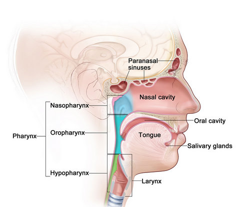
Image 1 : The most basic parts of throat anatomy.
Picture Source : www.ncbi.nlm.nih.gov
Gross Anatomy of throat
There are two major sections of the throat they are the pharynx and the larynx. These structures are shaped in a ring or a muscular tube.
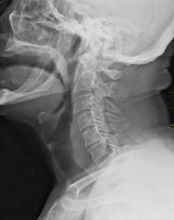
Picture 2 : The dark like at the front edge of the neck which shows where the throat is.
Picture Source : upload.wikimedia.org
Pharynx: Covers both the airway and the entrance to the digestive tract.
The nasopharynx starts at the back of the nasal cavity and ends at the soft palate. It contains the uvula which is a small piece of skin and fatty tissue which hangs at the back of the throat. (4,5, 6)
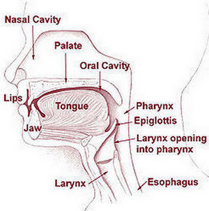 Figure 3 : The nasopharynx behind the oral cavity and above where the arrow is pointing to the pharynx.
Image Source : www.healthhype.com
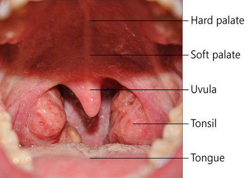
Picture 4 : A very clear image of the healthy tissue in the throat.
Image Source : www.healthhype.com
The oropharynx starts at the soft palate and ends at the hyoid bone. The oropharynx receives food bolus from the oral cavity through the oropharyngeal inlet. The inlet has palatoglossal muscles which create folds in the mucosa.
It also contains the vallecular or space between the base of the tongue and the epiglottis. Between the palatoglossal and palatopharyngeal folds of the oropharynx sit the palatine tonsils. The construction of food is done by the superior and middle pharyngeal constrictors.
These muscles have a mucous membrane covering them. This area also has a nerve which passes through it called the glossopharyngeal nerve. (1, 2, 5)
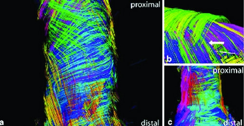
Image 5 : This image shows the dimensions of the muscle fibers in the esophagus.
Photo Source : www.ncbi.nlm.nih.gov
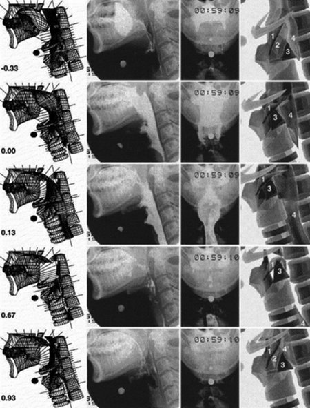
Figure 6 : This image was made using videofluoroscopy, It shows how the oropharyngeal area moves during a barium swallow test.
Picture Source : www.ncbi.nlm.nih.gov
The hypopharynx starts at the hyoid bone and ends at the upper esophageal sphincter. This area contains the epiglottis, the paired aryepiglottic folds, and the arytenoid cartilages.
The constricting muscles in this area are the middle and inferior constrictors which are also covered with an overlying mucous membrane.
Another important muscle in this area is the circumferential cricopharyngeus muscle which while resting is contracted and during swallowing it relaxes to let the food pass to the esophagus. (1, 2, 5)
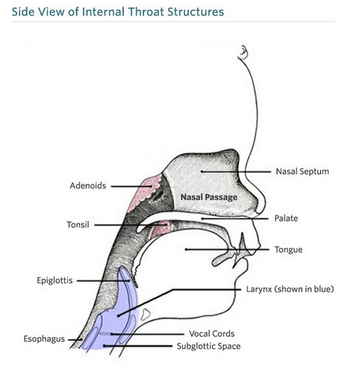 Picture 7 : This diagram shows the structures of the throat for a child.
Image Source : www.chop.edu
The larynx: It starts at the epiglottis and ends in the cricoid cartilage.
It is made up of many tissues such as cartilage muscle and other soft tissues. It contains the well-known folds which are called vocal cords. The vocal cords make a sound when air passes by them. These are not only for speech but also constrict to prevent choking or aspiration of food or objects into the trachea.
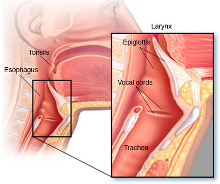
Image 8 : The folds that appear above the trachea called vocal folds or vocal cords.
Photo Source c: www.mayoclinic.org
Epiglottis- This is the part that closes the airway during swallowing. It is simply a piece of cartilage that is behind the tongue and at the start of the larynx. (1, 2, 5)
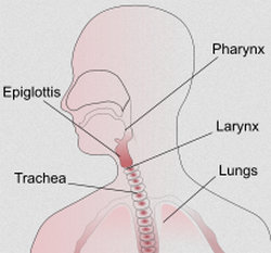
Picture 9 : The trachea is in comparison to the larynx and the lungs.
Image Source : upload.wikimedia.org
In this one of its most important functions is to protect the airway. It has various subdivisions which are:
Supraglottis – starts at the epiglottis and goes to the vocal folds
Glottis – starts at the level of the vocal folds
Subglottis – starts at the focal folds and goes to the corticoid cartilage. (1, 2)
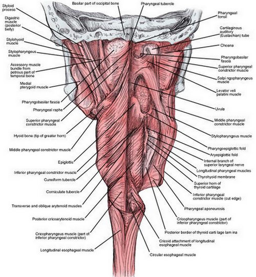
Figure 10 : This image shows the intensity of the muscular makeup of the throat.
Photo Source : img.medscapestatic.com

Image 11 : The complicated muscles in the pharynx
Picture Source : www.ncbi.nlm.nih.gov
On this web page, you can find a very interesting slide show of the neck which can show you various tissues that have been discussed in the article you just read.
http://www.healthline.com/human-body-maps/neck
In conclusion, the throat is complex and active. There are many tissues and structures that work together to assist in speech, breathing, and ignition of the digestive process.
References:
- http://emedicine.medscape.com/article/1899345-overview
- https://www.ncbi.nlm.nih.gov/pubmedhealth/PMHT0024473/
- https://en.wikipedia.org/wiki/Throat
- http://www.chop.edu/conditions-diseases/throat-anatomy-and-physiology
- http://www.throatproblems.co.uk/anatomy-throat.html
- http://www.healthhype.com/throat-anatomy-throat-parts.html
- http://diseasespictures.com/throat-anatomy-throat-parts-pictures-functions/
- http://www.medic8.com/healthguide/sore-throat/throat-anatomy.html
- https://www.ncbi.nlm.nih.gov/books/NBK54272/