Human Cell
Last reviewed by Dr. Raj MD on January 12th, 2022.
Every living thing has cells. It is the basic functional unit in humans. Humans are multi cellular organisms, which roughly have trillions of cells in the human body. They all play an important role in various bodily functions.
They form the body’s structure, take in and transport nutrients from foods to the host, convert nutrients to energy, and carry the genetic materials of the body. The cell, though extremely minute does have different parts on it. (1, 2)

Diagram 1: The anatomical presentation of the human cell.
Picture Source: www.printablediagram.com
How many cells are in the human body ?
Ans : Approx … 37.2 trillion cells
What are the different parts of the human cells? How do these parts function?
Cell membrane
It is the outer covering of the cell, which consists of proteins and lipids. It is semisolid, which aids in the movement of the cell organs to other places. It contains four molecules: carbohydrates, proteins phospholipids, and cholesterol.
The functions of the cell membranes are:
- It protects the cell.
- It regulates the exchange of substances in and out of the cell. (1, 2, 3)

Diagram 2: The human cell membrane.
Image Source: www.curezone.org
Cytoplasm/Protoplasm
It is the fluid inside the cells, which allow a number of cell organs to float inside the cell. It contains a nucleus surrounded by a nuclear membrane. It consists of molecules, enzymes, fatty acids, sugar, and amino acids. (3)
The functions of cytoplasm are:
- It contains molecules that aid in various metabolic functions of the body and facilitates the breakdown of waste materials.
- It gives the cell its shape.
- It keeps the organelle in place. (3, 4)
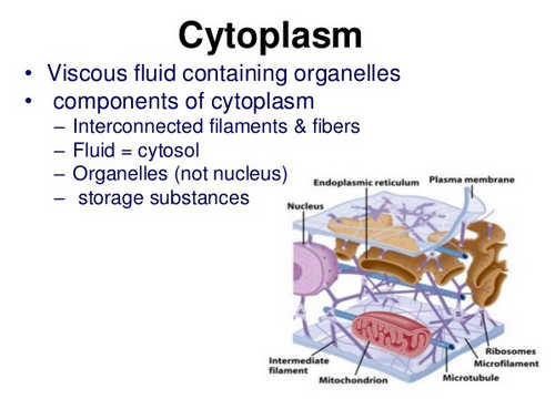
Image 3: The cytoplasm of the human cell.
Photo Source: image.slidesharecdn.com
Endoplasmic Reticulum
It consists of a network of membranes such as the tubules and vesicles. There are two kinds of endoplasmic reticulum: the rough and smooth. (4)
- Rough endoplasmic reticulum – disk-shaped/shape of sheets. The ribosome is attached to the surface of the rough endoplasmic reticulum.
- Smooth endoplasmic reticulum – A tube-like structure, which acts as the storehouse of steroids and lipids. It has a special structure called sarcoplasmic reticulum that stores different types of ions which can be used by the body in times of emergency. (4, 5)
The functions of the endoplasmic reticulum are:
- It is the manufacturing and packaging unit of the cell.
- It temporarily isolates one organ from the other until the manufacturing process is complete.
- It folds and transports various types of proteins. (5, 6)
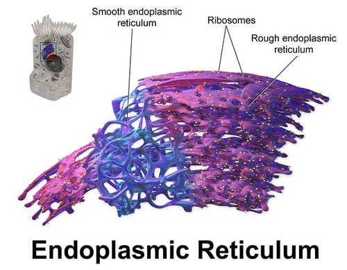
Photo 4: A closer look at endoplasmic reticulum.
Diagram Source: study.com
Lysosomes
They are vesicles that break off from the Golgi bodies. They contain enzymes responsible for digestion of nutrients in the cell. The functions of lysosomes are as follows:
- It is the garbage disposal center of the cell. Lysosomes break down leftover cellular waste.
- It is called the suicide bag of the cell because it destroys leftover content.
- It is the one in charge of cellular homeostasis. It also plays an important role in the repair of plasma membrane, metabolism of energy, and cell signaling functions.
- It protects the cells from diseases. (5, 6, 7, 8)
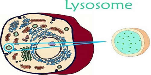
Picture 5: The lysosome of the human cell.
Photo Source: www.assignmentpoint.com
Peroxisomes
They are similar to lysosomes. They contain enzymes that combine together to form hydrogen peroxide, which neutralizes toxic substances in the cell. The functions of peroxisomes are:
- They keep the liver healthy.
- They metabolize energy and hold digestive enzymes that break down toxic materials in the cells. (7, 8)
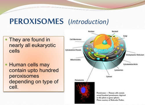
Diagram 6: A human cell contains up to a hundred peroxisome.
Picture Source: image.slidesharecdn.com
Golgi Bodies/Golgi Apparatus
It consists of membrane-bound sacs called cisternae. A single Golgi apparatus contain somewhere between five to eight cisternae. The role of Golgi bodies are:
- They process and bundle macromolecules in the cells.
- They modify, sort, and package proteins. That’s why Golgi bodies are tagged as the post office of the cell. They gather simple molecules and group them to form a complex.
- They play an important role in building lysosomes, which are used for digestion of nutrients. (8, 9, 10)
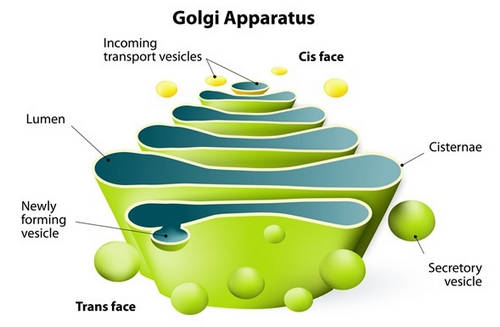
Picture 7: The Golgi apparatus of the human cell.
Diagram Source: www.news-medical.net
Mitochondria/Mitochondrion
A mitochondrion is a small structure inside the cell, which has two membranes and a matrix. Chemical reactions occur in the membrane and the fluid is held in the matrix. It is the powerhouse of the cell because it supplies continuous energy to the cell. The number of mitochondria varies from one cell to another. Some have thousands while others have none. The number of mitochondria is determined by the energy requirement. A cell that needs more energy has more mitochondria than a cell that does not need much energy.
The functions of mitochondria are as follows:
- They are responsible for cellular respiration (breakdown of nutrients and giving the energy needed by the cell).
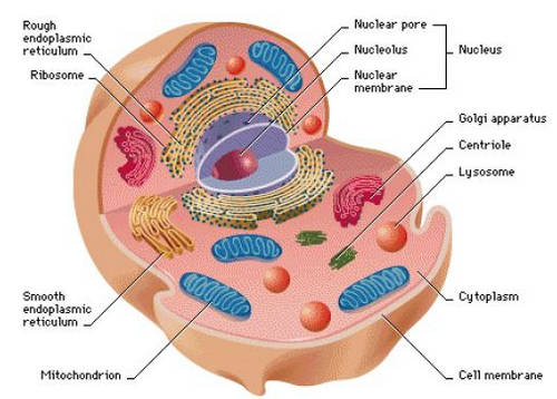
Diagram 8: The mitochondria of the human cell.
Picture Source: justmeint1health.files.wordpress.com
Nucleus
It contains the genetic material of the cell that is organized as DNA molecules and a variety of proteins to form chromosomes. The nucleus has double membrane nuclear envelope encasing the entire organelle. It isolates its content from the rest of the cell.
The functions of the nucleus include the following:
- It contains the genes and facilitates DNA replication during the cell cycle.
- It controls the overall function of the cell making it the control center or the brain of the cell.
- It controls various processes such as eating, reproduction, and movement of the cells.
- The anatomy and physiology of the human body are determined by the nucleus. (2, 6, 8)
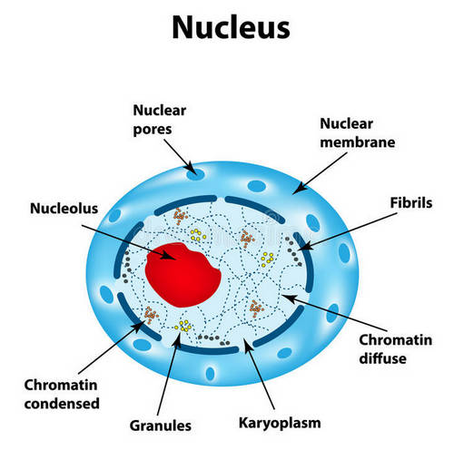
Diagram 9: The nucleus of the human cell.
Picture Source: thumbs.dreamstime.com
Microfilaments
A microfilament, also known as actin filament is a thin protein structure in the human cell. Although microfilaments are thin, they are strong and flexible enough to hold the cell’s shape and facilitate cellular movement.
The functions of microfilaments are:
- They hold the cells’ shape.
- They facilitate movement of the cell.
- They play a vital role in the contraction of muscle tissues. (2, 3, 4)
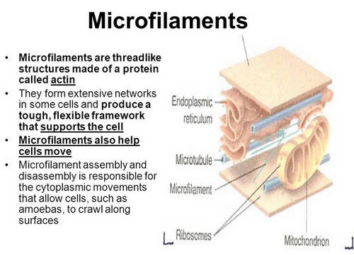
Diagram 10: A microfilament is a structure in the cell that looks like a thread.
Photo Source: slideplayer.com
Microtubules
Microtubules are moving chromosomes. They are hollow fibrous shafts.
The functions of microtubules are:
- They are essential for cell division. Microtubules pair with chromosomes enabling the chromosomes to split and attach to new daughter cell.
- Microtubules support and give cells its shape.
- They help transport materials to and from the cells.
- They form a large structure outside the cells.
- They can combine in specific bundles forming cilia and flagella, which play a very important role in the cellular movement. (4, 7, 9)
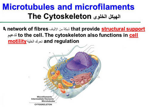
Image 11: The microtubules of the human cell.
Diagram Source: images.slideplayer.com
For more info on Human cell atlas – https://www.humancellatlas.org/
References:
- https://ghr.nlm.nih.gov/primer/basics/cell
- http://diseasespictures.com/human-cell-functions-diagram-parts-pictures-structure/
- http://www.healthhype.com/human-cell-diagram-parts-pictures-structure-and-functions.html
- https://www.visiblebody.com/blog/anatomy-and-physiology-parts-of-a-human-cell
- http://www.medibiztv.com/articles/brief-introductio-on-human-cell-structure-and-its-functions
- http://people.eku.edu/ritchisong/301notes1.htm
- https://www.ck12.org/section/Cell-Parts-and-Their-Functions/
- http://www.biologyguide.net/resources/bk/cell_structure_function.php
- https://web.sonoma.edu/users/c/cannon/110chapter4.html
- http://www.juniordentist.com/cell-components-and-functions-of-cell-organelles.html